South Australian Medical Heritage Society Inc
Website for the Virtual Museum
Home
Coming meetings
Past meetings
About the Society
Main Galleries
Medicine
Surgery
Anaesthesia
X-rays
Hospitals,other organisations
Individuals of note
Small Galleries
Ethnic medicine
- Aboriginal
- Chinese
- Mediterran
Photodynamic Therapy
[A recent editorial on Photodynamic therapy and cancer may be found in the BMJ 2009;339:b2459]
ACKNOWLEDGMENTS: Jan Hooper, Director of Nursing (Medical), kindly allowed us to take photographs of the now outdated Photo Cure ASA machine. Dr. Warren Weightman, head of the Dermatology Department at TQEH, provided the information and photograph of a current model.
The concept of Photodynamic therapy has been known for a long time. At the beginning of the 20thcentury Neils Finsen and Oscar Raab noted that haemato-porphyrin had a tumour localising tendency. They also noted that if the tumour was irradiated with red light (>610 nm) it produced a photo-toxic effect on the tumour cells. In the last 25 years this method of treatment became more popular and is now expanding into all fields of medicine and surgery.
The literature reports by the proponents of this method of treatment claim a success rate similar to cryotherapy and surgery when used in the treatment of hyper-keratoses, basal cell skin cancers and other suitable lesions.
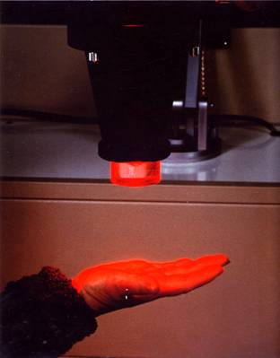
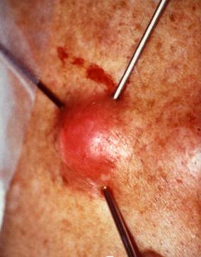
In South Australia during the early 1990s Dr. Ian Forbes then a Reader in Medicine at the Queen Elizabeth Hospital in Adelaide used intravenous injection of a haemato-porphyrin derivative (HpD). The haemato-porphyrin was prepared in the University Department of Chemistry, and the Pharmacy Department of the Hospital provided the required sterile conditions. It was preferentially concentrated in the skin lesions and after irradiation with a red light source, with a wavelength of 620 nanometers (nm), achieved several cures. In one instance he treated local recurrences (small nodules) after mastectomy and achieved a cure (personal communication Michael Patkin). The associated transient photosensitivity however was troublesome.
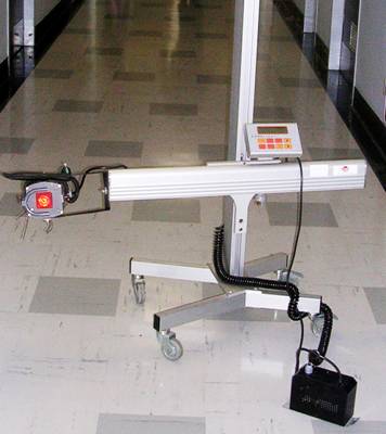
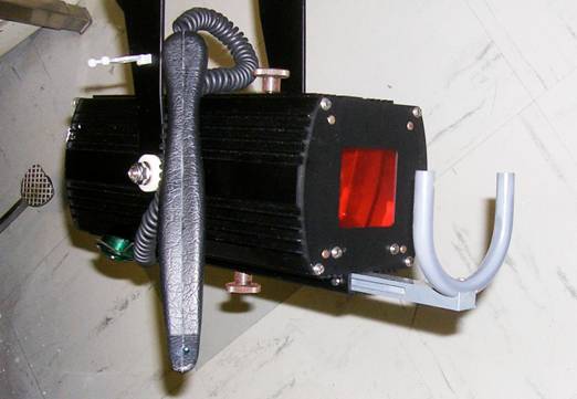
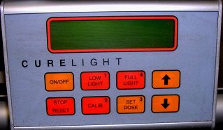
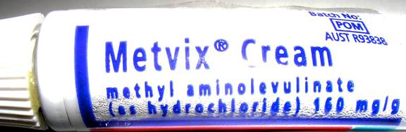
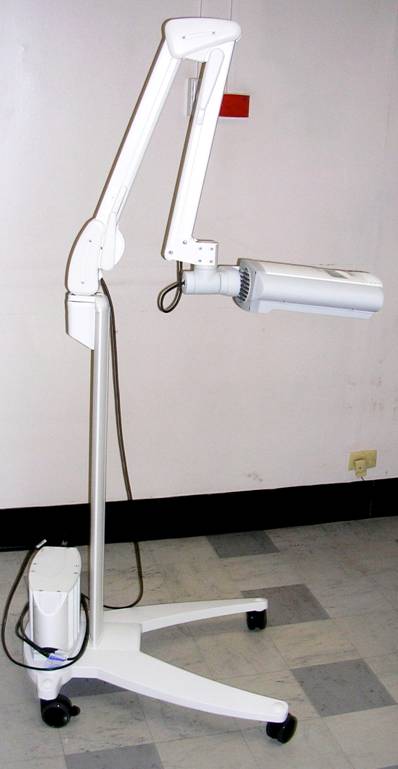
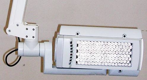
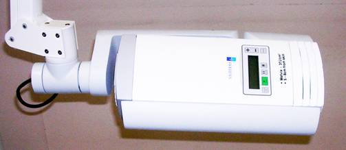
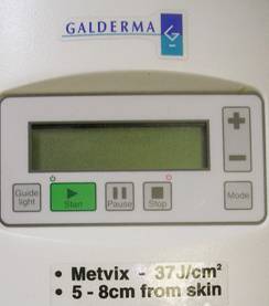
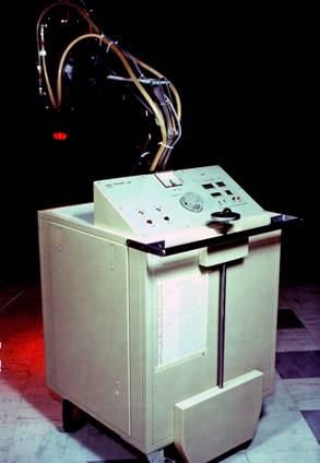
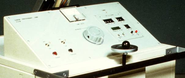
The QEH Dermatology Department later purchased a PhotoCure ASA machine from Norway for the same reason. In addition the company also supplied a cream ("Metvix"). When applied directly to the lesion this confined the photo-sensitivity to the local area only. It was used with success in selected cases and when the company produced a new more efficient model the old machine was replaced.
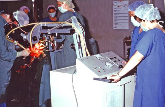
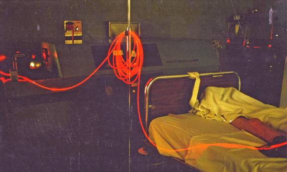
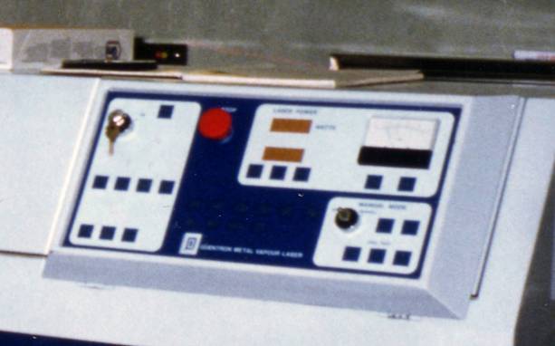
Later, a gold laser was used. This provided a red light at 627.8 nm. And was developed by Quentron Optics, a South Australian company. In some instances the results were spectacular but the therapy had little effect on advanced or large tumours. In addition several treatments and occasional anaesthetics were needed and following photosensitivity was troublesome.
More recently the combination of photo-sensitising agent and illumination is also used for diagnostic purposes. An agent ("Hexvix") is injected intravenously. Because it as preferentially concentrated in a polyp or tumour it fluoresces with an appropriate light source. It is claimed that this type of imaging improves the accuracy of diagnosis of bladder, colon, or bronchial tumours and has treatment possibilities. The Company logo of the early model is shown below.

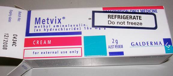
Metvix cream container and tube (below).
Distributed by the Australian branch of the Swiss firm Galderma.
Addendum
ACKNOWLEDGMENT: The additional following photographs have been provided by Dr. Prue Cowled, Principal Scientist in the Department of Surgery at The Queen Elizabeth Hospital, Adelaide . The details and information has been kindly supplied by Dr. Ian Forbes
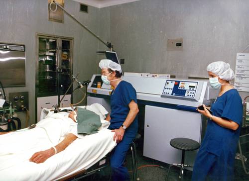
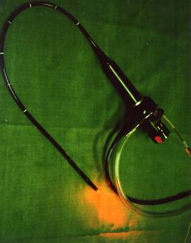
bronchoscope ....
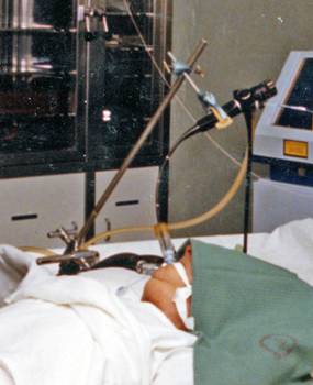 ..... under general anaesthetic. Fibre-optic cable is passed through the biopsy channel of the bronchoscope to illuminate the tumour. Bronchoscope held by clamp.
-o0o-